Home
/ Loculated Pleural Effusion Cxr : Pleural Effusion : But clinical identification of pleural effusion is possible only when the amount of fluid is more than 500ml.
Loculated Pleural Effusion Cxr : Pleural Effusion : But clinical identification of pleural effusion is possible only when the amount of fluid is more than 500ml.
Loculated Pleural Effusion Cxr : Pleural Effusion : But clinical identification of pleural effusion is possible only when the amount of fluid is more than 500ml.. Cxr/ct chest demonstrated right sided pleural effusion. The first step in evaluating pleural effusions is determining whether it is transudative or exudative. A chest tube (12f) was inserted under imaging guidance into the largest locule. Expressed as 29.5% (23.3) vs. This chapter describes the usual causes (lung cancer, breast cancer, lymphoma, mesothelioma), clinical features, imaging, and management of malignant pleural effusions, parapneumonic effusions, empyema, tuberculous effusions, as well as rarer causes.
It detects pleural effusions with higher sensitivity and specificity than cxr, and provides valuable information about the size and depth of the pleural effusion, the echogenicity of the fluid, the presence of septated or loculated fluid, pleural thickening and nodularity, and the presence of any contralateral pleural effusion. In chf effusions are bilateral and more on right. In contrast, tpa or dnase alone did not improve radiographic clearance. In a pleural effusion, whether mobile (fig. The fluid buildup may be the result of a chronic condition like congestive heart failure.
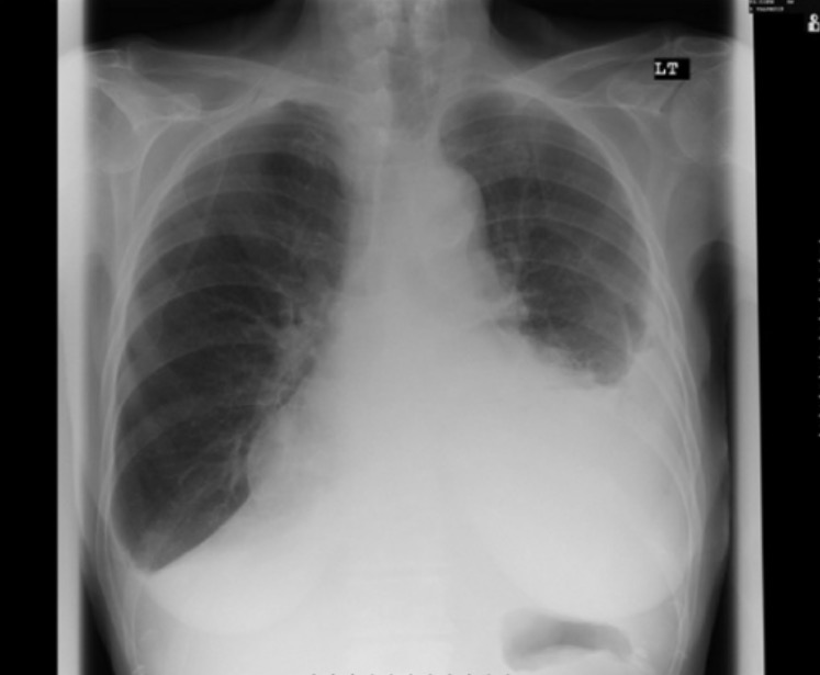
Pleural Effusion Concise Medical Knowledge from cdn.lecturio.com 6.9b), the lung is intact but displaced away from the abnormality. Loculated right pleural effusion with foci of atelectasis and consolidative changes concerning for pneumonia. All patients were subjected to routine chest radiography (cxr; But clinical identification of pleural effusion is possible only when the amount of fluid is more than 500ml. Loculation most commonly occurs with exudative fluid, blood and pus. Recent research into the causes and management of pleural effusion has altered clinical practice. 1 article features images from this case 20 public playlist includes this case Optimal candidates for pigtail catheters are those with thin, free flowing fluids such as air or new pleural effusions
An empyema, by definition, is frank pus.
I a chest radiograph appropriate for age An empyema, by definition, is frank pus. A pleural effusion is a collection of fluid in the pleural space. But, catheter removal is suggested if the infection fails to improve. Most effusions start like this and can be easily missed. All patients were subjected to routine chest radiography (cxr; What are loculated pleural effusions? There is a large left pleural effusion obscuring the lower half of the left hemi thorax. Depending on the clinical context, ultrasonography or computed tomography (ct) scanning can be used to confirm a pleural effusion, especially in cases of loculated pleural effusion, complete. • in patients with symptomatic mpes with nonexpandable lung, failed pleurodesis, or loculated effusion, ipcs are suggested over chemical pleurodesis. Pleural effusion is an abnormal accumulation of fluid in the pleural space. Loculation most commonly occurs with exudative fluid, blood and pus. Pleural fluid glucose < 60 mg/dl;
The lack of specificity is mainly due to the limitations of the imaging modality. Depending on the clinical context, ultrasonography or computed tomography (ct) scanning can be used to confirm a pleural effusion, especially in cases of loculated pleural effusion, complete. Optimal candidates for pigtail catheters are those with thin, free flowing fluids such as air or new pleural effusions 17.2% (19.6) of the hemithorax on cxr. Most effusions start like this and can be easily missed.
Intrapleural Urokinase For The Treatment Of Loculated Malignant Pleural Effusions And Trapped Lungs In Medically Inoperable Cancer Patients Journal Of Thoracic Oncology from els-jbs-prod-cdn.jbs.elsevierhealth.com A complicated parapneumonic effusion has the following biochemical, microbiologic, and anatomic features: Most pleural effusions, whether free flowing or loculated, are hypoechoic with a sharp echogenic line that delineates the visceral pleura and lung. But, catheter removal is suggested if the infection fails to improve. Sided pleural effusion that appears loculated with a right lower lobe opacification concerning for compressive atelectasis versus consolidation. Ph < 7.2, lactate dehydrogenase (ldh) > 1,000 u, gram stain or culture positive, and loculations and/or septations. The primary outcome was the absolute change in the pleural opacity on a frontal chest radiograph between days 1 and 7. Ph 6.09, lactate dehydrogenase 71,300 u/l, protein 40 g/l but no microorganism was cultured. A chest tube (12f) was inserted under imaging guidance into the largest locule.
17.2% (19.6) of the hemithorax on cxr.
She was coagulopathic (activated partial thromboplastin time (aptt) of 44.4 s and international normalized ratio (inr) of 1.8). Lateral decubitus views are frequently necessary for distinguishing free pleural effusion from pleural thickening, but loculated effusions are not as easily distinguished from pleural thickening. Loculated right pleural effusion with foci of atelectasis and consolidative changes concerning for pneumonia. 6.9b), the lung is intact but displaced away from the abnormality. An empyema, by definition, is frank pus. 3 the mortality rate from pleural infection is estimated to be between 10% and 20%. Ray, and after treatment (ie drainage), there should be a difference, however, if a cxr is taken day/ month. Most pleural effusions, whether free flowing or loculated, are hypoechoic with a sharp echogenic line that delineates the visceral pleura and lung. A pleural effusion is a collection of fluid in the pleural space. Fluid gathers in the lowest part of the chest, according to the patient's position. There is a large left pleural effusion obscuring the lower half of the left hemi thorax. Differential diagnosis of pleural effusion; Sometimes in the setting of pleuritis, loculation of fluid may occur within the fissures or between the pleural layers (visceral and parietal).
In a pleural effusion, whether mobile (fig. So pleural effusion is seen on a chest x. Loculated right pleural effusion with foci of atelectasis and consolidative changes concerning for pneumonia. Ph < 7.2, lactate dehydrogenase (ldh) > 1,000 u, gram stain or culture positive, and loculations and/or septations. Most effusions start like this and can be easily missed.
Loculated Pleural Effusion On Cxr Radiology Case Radiopaedia Org from prod-images-static.radiopaedia.org But, catheter removal is suggested if the infection fails to improve. Transudative effusions are a result of pressure filtration without capillary injury (i.e hydrostatic and oncotic pressure abnormalities). A pleural effusion is a collection of fluid in the pleural space. So pleural effusion is seen on a chest x. The fluid buildup may be the result of a chronic condition like congestive heart failure. A 14 french pigtail catheter was placed with successful drainage of the pleural effusion and improvement of respiratory rate/o2sat. I a chest radiograph appropriate for age All patients were subjected to routine chest radiography (cxr;
• in patients with symptomatic mpes with nonexpandable lung, failed pleurodesis, or loculated effusion, ipcs are suggested over chemical pleurodesis.
The first step in evaluating pleural effusions is determining whether it is transudative or exudative. 6.9b), the lung is intact but displaced away from the abnormality. But clinical identification of pleural effusion is possible only when the amount of fluid is more than 500ml. Most effusions start like this and can be easily missed. In endobronchial obstruction, the air distal to the point of obstruction is resorbed—a postobstructive atelectasis results (fig. Loculated right pleural effusion with foci of atelectasis and consolidative changes concerning for pneumonia. This chapter describes the usual causes (lung cancer, breast cancer, lymphoma, mesothelioma), clinical features, imaging, and management of malignant pleural effusions, parapneumonic effusions, empyema, tuberculous effusions, as well as rarer causes. A chest tube (12f) was inserted under imaging guidance into the largest locule. Like pleural effusion, pleural thickening is usually appreciated as a thick white line between the lucent lungs and ribs. All patients were subjected to routine chest radiography (cxr; There is an increasing role for ct and mri. Cxr/ct chest demonstrated right sided pleural effusion. Ph < 7.2, lactate dehydrogenase (ldh) > 1,000 u, gram stain or culture positive, and loculations and/or septations.
Read more 3 doctors agree loculated pleural effusion. It detects pleural effusions with higher sensitivity and specificity than cxr, and provides valuable information about the size and depth of the pleural effusion, the echogenicity of the fluid, the presence of septated or loculated fluid, pleural thickening and nodularity, and the presence of any contralateral pleural effusion.